Wnt is a hybrid name derived from the combination of the mouse proto-oncogene int-1 and the segment-polarity gene wingless (wg) from Drosophila, which form the Wnt gene family since a revision of the nomenclature in the early 1990s1. The evolutionary-conserved Wnt proteins have been implicated in the regulation of cell proliferation and development through several different signal-transduction pathways. Through its numerous interactions, perturbations in these pathways lead to numerous defects such as cancer (reviewed in 2,3) and degenerative diseases (reviewed in 4).
The canonical Wnt or Wnt/β-catenin signaling pathway (see figure) begins with the binding of Wnt to a receptor complex consisting of frizzled (Fz) and LRP (lipoprotein receptor-related protein). After activation of the complex, Fz interacts with disheveled (Dsh) in the cytosol, which causes the aggregation of a complex consisting of Axin, GSK-3β, CK1α, and APC at the receptor. In this complex, glycogen synthase kinase 3β (GSK-3ß) is inactivated and prevents the transcriptional cofactor β-catenin from being phosphorylated. Increased levels of cytosolic β-catenin allow translocation to the nucleus, removing the suppression of gene transcription by the TCF/LEF complex.
In its inactive configuration, Axin-bound β-catenin is inactivated due to its phosphorylation by serine-threonine kinases CK1α and GSK-3ß and is subsequently destroyed by ubiquitin-mediated proteolysis. This prevents β-catenin from entering the nucleus and inactivating the repressor complex consisting of TCF (T-cell specific factor) and LEF (lymphoid enhancer-binding factor), which prevents gene transcription of the target genes.
The non-canonical or β-catenin independent Wnt signaling pathway uses other effectors to regulate transcription. One of them is the Wnt/Ca2+ signaling pathway, whose regulation relies on the transcription factors NFAT (nuclear factor of activated T cells) and TAK1-induced Nemo-like kinase (NLK) and is involved in cancer, inflammation, and neurodegenerative diseases. Additionally, the PCP signaling pathway activates a number of Rho family of GTPases as well as Jun-N-terminal kinase (JNK) and regulates cell polarity during morphogenesis.
The most important question in the exploitation of the Wnt pathway to combat disease is how to control aberrations without interfering with the normal functions of this complex pathway. To foster Wnt signaling research, Rockland is providing several specific antibodies to Wnt signaling modulators.
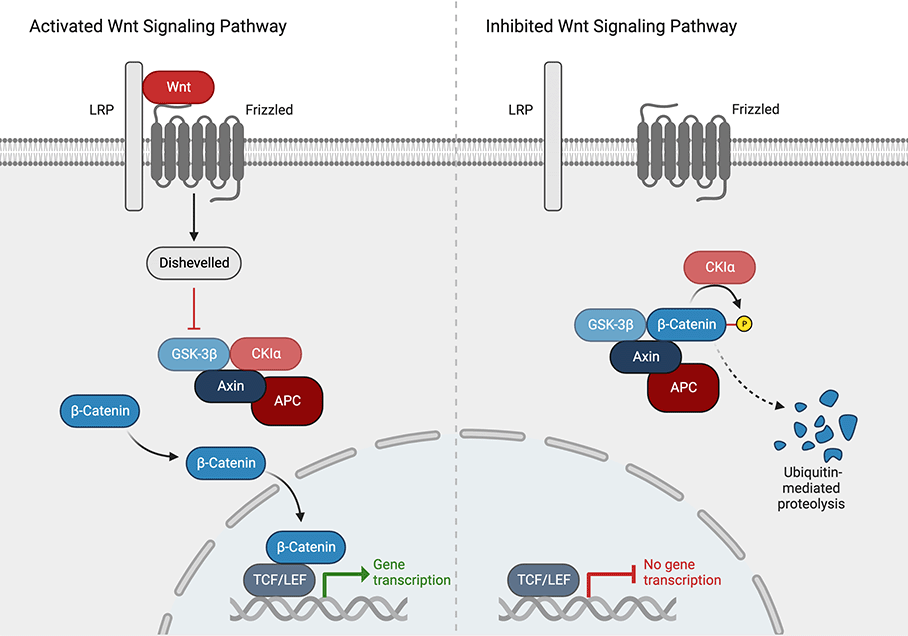
Figure: Canonical Wnt or Wnt/β-catenin signaling pathway. Left: activated pathway by binding of Wnt to the receptor complex consisting of frizzled and LRP. Right: inhibited pathway by phosphorylation of β-catenin and subsequent degradation. (Adapted from Zhang & Wang, 2020, created with BioRender.com)
Wnt Pathway Antibodies
| Product | Clonality | Reactivity | Applications |
| AXIN1 Antibody | Polyclonal | Human, Mouse, Rat | WB, IF, ELISA |
| AXIN2 Antibody | Polyclonal | Human, Mouse, Rat | WB, IHC, IF, ELISA |
| BAMBI Antibody | Polyclonal | Human, Mouse, Rat | WB, IHC, IF, ELISA |
| beta Catenin Antibody | Polyclonal | Zebrafish, Human | WB, IHC, IF, ELISA |
| Beta TrCP2 Antibody | Polyclonal | Human, Mouse | WB, ELISA |
| CaMKII Antibody | Monoclonal | Bovine, Mouse, Rat | WB, IHC, IF, IP |
| CaMKII Antibody | Monoclonal | Rat | WB, IHC, IF, IP |
| CaM Kinase II Antibody | Polyclonal | Mouse | WB, ELISA |
| CaM Kinase II phospho T286 Antibody | Polyclonal | Mouse, Rat | WB |
| CREBBP Antibody | Polyclonal | Human, Mouse | WB, ELISA |
| CXXC4 Antibody | Polyclonal | Human, Mouse, Rat | WB, IHC, IF, ELISA |
| GSK3 Alpha Antibody | Polyclonal | Human | WB, ELISA |
| GSK3 Alpha phospho S21 Antibody | Polyclonal | Human | WB, ELISA |
| GSK3 Beta phospho S9 Antibody | Polyclonal | Human | WB, IHC, ELISA |
| LGR4 Antibody | Monoclonal | Human | WB, ELISA |
| MAPK 8/9 Antibody | Polyclonal | Human, Mouse, Rat | WB, IHC |
| NFATc1 Antibody | Polyclonal | Human, Mouse | WB, IF |
| NOTUM Antibody | Polyclonal | Human, Mouse, Rat | WB, IHC, IF, ELISA |
| Nuclear receptor ROR gamma phospho S203 Antibody | Polyclonal | Mouse | WB, ELISA |
| Presenilin1 Antibody | Polyclonal | Human, Mouse, Rat | WB, IHC, IF, ELISA |
| PRICKLE1 Antibody | Polyclonal | Human, Mouse, Rat | WB, IF, ELISA |
| Protein Kinase C Beta Antibody | Polyclonal | Human | WB, ELISA |
| Protein Kinase C delta-Binding Protein Antibody | Polyclonal | Human | IHC |
| Rock-2 phospho Y256 Antibody | Polyclonal | Human | WB, ELISA |
| RSPO1 Antibody | Polyclonal | Human, Mouse, Rat | WB, IHC, IF, ELISA |
| SOX17 Antibody | Polyclonal | Human, Mouse, Rat | WB, IF, ELISA |
| SMAD3 Antibody | Polyclonal | Human, Mouse | WB, IHC, ELISA |
| SMAD3 Antibody | Polyclonal | Human, Mouse, Rat | WB, ELISA |
| SMAD3 Antibody | Polyclonal | Human | WB, ELISA |
| SMAD3 phospho S423/phospho S425 Antibody | Polyclonal | Human | WB, IHC, IF, IP, ELISA |
| SMAD3 phospho T179 Antibody | Polyclonal | Mouse | WB, IP, ELISA |
| SMAD4 Antibody | Polyclonal | Human, Mouse, Xenopus | WB, ELISA |
| SUMO Antibody | Polyclonal | Human | WB |
| TAK1 Antibody | Polyclonal | Human, Mouse, Rat | WB, IF, ELISA |
| Wnt1 Antibody | Polyclonal | Human, Mouse | WB, ELISA |
| Wnt1 Antibody | Monoclonal | Mouse | WB |
| Wnt5A Antibody | Polyclonal | Human, Mouse, Rat | WB, IHC |
| Wnt10a Antibody | Polyclonal | Human, Mouse, Rat | WB, IHC, IF, ELISA |
| Wnt10B Antibody | Polyclonal | Human, Mouse, Rat | WB, IHC, ELISA |
| ZBED3 Antibody | Polyclonal | Human | WB, IHC, ELISA |
| ZBED3 Antibody | Polyclonal | Human, Mouse, Rat | WB, ELISA |
Wnt Pathway ELISA Kits
| Product | Application |
| Human DKK-1 ELISA Kit | ELISA |
| Mouse DKK1 ELISA Kit | ELISA |
| Rat DKK1 ELISA Kit | ELISA |
| Human DKK-3 ELISA Kit | ELISA |
| Mouse Sclerostin - SOST ELISA Kit | ELISA |
| Human WISP1 - CCN4 ELISA Kit | ELISA |
| Mouse WISP1 - CCN4 ELISA Kit | ELISA |
References
- Nusse, R., Brown, A., Papkoff, J., Scambler, P., Shackleford, G., McMahon, A., Moon, R., & Varmus, H. (1991). A new nomenclature for int-1 and related genes: the Wnt gene family. Cell, 64(2), 231.
- Patel, S., Alam, A., Pant, R., & Chattopadhyay, S. (2019). Wnt Signaling and Its Significance Within the Tumor Microenvironment: Novel Therapeutic Insights. Frontiers in immunology, 10, 2872.
- Zhang, Y., & Wang, X. (2020). Targeting the Wnt/β-catenin signaling pathway in cancer. Journal of hematology & oncology, 13(1), 165.
- Nusse, R., & Clevers, H. (2017). Wnt/β-Catenin Signaling, Disease, and Emerging Therapeutic Modalities. Cell, 169(6), 985–999.