Red fluorescent proteins (RFPs) have become essential tools in molecular and cell biology for tracking gene expression, lineage tracing, and protein localization in living systems. First identified in Discosoma species, RFPs expanded the fluorescent protein toolkit beyond Green Fluorescent Protein by enabling multi-color imaging and more complex experimental design.
Like GFP, RFPs form an internal chromophore through autocatalytic cyclization of amino acids within a β-barrel structure. However, early RFPs such as DsRed presented practical limitations, including slow maturation and obligate tetramerization, which could interfere with protein function when used as fusion tags. These challenges led to the development of a wide range of engineered RFP variants optimized for brightness, maturation time, and monomeric behavior.
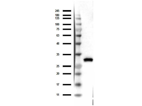
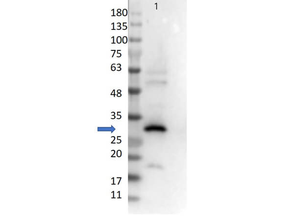
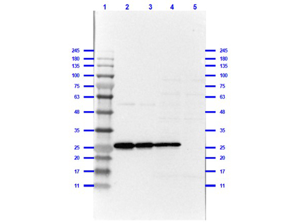
In many experimental workflows, including Western blotting, immunohistochemistry, and fixed-cell imaging, the intrinsic fluorescence of RFP is not sufficient or is lost entirely. In these cases, anti-RFP antibodies are required for sensitive and reliable detection of tagged proteins. Rockland anti-RFP antibodies are designed to provide consistent detection across experimental conditions and are widely cited in literature.
RFP Variants: From DsRed to mScarlet
The evolution of RFPs from tetrameric DsRed to modern monomeric variants represents a major advancement in fluorescent protein engineering. DsRed2 improved maturation and reduced cytotoxicity but retained oligomerization. Subsequent variants addressed these limitations more effectively.
mCherry, derived through directed evolution, is a true monomer with rapid maturation and good performance in denaturing conditions, making it suitable for biochemical assays such as Western blotting. tdTomato, a tandem dimer, offers exceptional brightness and is widely used in lineage tracing models. More recent variants, such as mScarlet-I, provide improved quantum yield and brightness for imaging applications, while others such as mOrange, mStrawberry, and mPlum expand the spectral range for multiplex experiments.
Although these variants differ in optical and biochemical properties, they retain conserved structural elements. Rockland polyclonal anti-RFP antibodies are raised against full-length DsRed and recognize epitopes shared across the RFP family, enabling detection of multiple variants, including mCherry, tdTomato, mOrange, and related proteins, all with a single reagent.
Importantly, RFPs share limited sequence identity with GFP (~19%), and anti-RFP antibodies do not cross-react with GFP variants. This allows for clean signal separation in dual-reporter experiments when used alongside anti-GFP antibodies.
| Variant | Ex (nm) | Em (nm) | Extinct. Coeff. (M-1cm-1) | Quantum yield (Φ) | Brightness (nM-1cm-1) | Maturation |
| DsRed | 558 | 583 | 72,500 | 0.68 | 49.3 | 24+ hours |
| DsRed2 | 561 | 587 | 52,000 | 0.55 | 28.6 | 6.5 hours |
| tdTomato | 554 | 581 | 138,000 | 0.69 | 95.22 | 1 hour |
| mCherry | 587 | 610 | 72,000 | 0.22 | 15.84 | 15 min |
| mOrange | 548 | 562 | 71,000 | 0.69 | 48.99 | 2.5 hours |
| mStrawberry | 574 | 596 | 90,000 | 0.29 | 26.1 | 50 min |
| mBanana | 540 | 553 | 6,000 | 0.7 | 4.2 | 1 hour |
| mPlum | 590 | 649 | 41,000 | 0.1 | 4.1 | 1.5 hours |
Choosing a Host Species for Your Anti-RFP Antibody
Anti-RFP antibodies are available from several host species, including rabbit, goat, mouse, chicken, and llama. Differences between these antibodies arise primarily from their structure and immunological properties, which influence their performance in specific applications.
| Host Species | Ig Class | Clonality | Key Advantages | Applications |
| Rabbit | IgG | Polyclonal | High affinity and sensitivity; multi-epitope binding across RFP variants; pre-adsorbed format minimizes background against human, mouse, and rat serum proteins; well-established secondary antibody ecosystem. | IF, IHC, WB, FC, IP, EM, FISH, multiplex |
| Goat | IgG | Polyclonal | Orthogonal host to rabbit, enables simultaneous use of a rabbit primary against a second target in the same section without secondary cross-reactivity; broad Ig repertoire; well-suited to dual-label multiplex workflows. | WB, IF, IHC, FC, EM, multiplex |
| Mouse | IgG | Monoclonal | Single defined epitope; exceptional lot-to-lot consistency; eliminates batch variability across long-term studies; confirmed reactivity across mCherry, tdTomato, mOrange, mStrawberry, mBanana, mPlum, and DsRed variants. | WB, ELISA, IF, IHC, FC, FISH, multiplex |
| Chicken | IgY | Polyclonal | IgY does not interact with mammalian Fc receptors or complement, eliminates non-specific background in tissues rich in Fc receptors (spleen, liver, lymph nodes); structurally distinct from mammalian IgG enabling true tri-label multiplex with rabbit and goat antibodies; harvested from egg yolk (non-terminal collection). | IF, IHC, FC, WB, multiplex; especially valuable in Fc receptor-rich tissue types and three-color panels alongside rabbit and goat primaries. |
| Llama | VHH | Recombinant Monoclonal | Single-domain antibody (~15 kDa); accesses epitopes sterically blocked from conventional antibodies; no Fc region eliminates all Fc receptor interactions; bacterially expressed for high reproducibility; easily conjugated for direct detection. | WB, ELISA, IF, FC; ideal for crowded antigen environments, proximity ligation assays, and workflows requiring small probe size or direct labeling without secondary antibodies. |
Conclusion
RFP antibodies are essential tools for the reliable detection of fluorescent protein reporters across a wide range of experimental conditions. The diversity of RFP variants, combined with differences in assay requirements, makes antibody selection an important consideration in experimental design.
Rockland anti-RFP antibodies are developed to recognize conserved epitopes across multiple RFP variants, providing broad applicability without the need for variant-specific reagents. With options across multiple host species and both monoclonal and polyclonal formats, these antibodies support applications ranging from Western blotting to multiplex immunofluorescence.
Selecting the appropriate antibody depends on the specific experimental context, including the RFP variant, the application, and the overall panel design. Product-specific validation data and literature references remain key resources for making an informed choice.
Featured Rockland Anti-RFP Publications
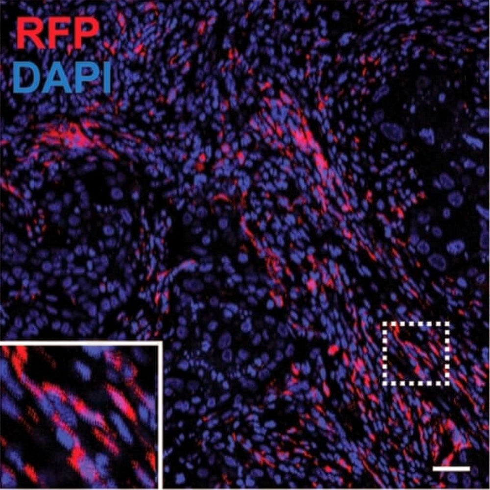
Lineage Tracing Reveals the Origins and Dynamics of Macrophages in Lung Injury and Repair
Jin, Mou, Zhu et al. Cell Discovery (Nature Portfolio), 2026.
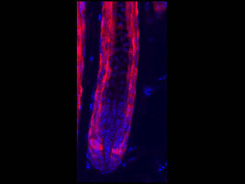
PPARγ Mediates Transdifferentiation of CX3CR1⁺-Derived Cells into Adipocytes
Yang, Y.-F.; Ruan, C.-C.; Lei, Y. Int. J. Mol. Sci., 2026.
TRACR: An Anterograde Transneuronal Tracing System for Genetic Access Across Synapses and Longitudinal Circuit Analysis
Lischinsky et al. bioRxiv (preprint), 2026.
Recommended RFP Antibodies
References
- Matz MV et al. Fluorescent proteins from nonbioluminescent Anthozoa species. Nature Biotechnology. 1999;17:969–973.
- Shaner NC et al. Improved monomeric red, orange and yellow fluorescent proteins derived from Discosoma sp. red fluorescent protein. Nature Biotechnology. 2004;22:1567–1572.
- Bindels DS et al. mScarlet: a bright monomeric red fluorescent protein for cellular imaging. Nature Methods. 2017;14:53–56.
- Lambert TJ. FPbase: a community-editable fluorescent protein database. Nature Methods. 2019;16:277–278.
- Shaner NC et al. A guide to choosing fluorescent proteins. Nature Methods. 2005;2:905–909.
- Jin H, Mou J, Zhu H et al. Lineage tracing reveals the origins and dynamics of macrophages in lung injury and repair. Cell Discovery. 2026;12:3.
- Yang YF, Ruan CC, Lei Y. PPARγ Mediates Transdifferentiation of CX3CR1⁺-Derived Cells into Adipocytes. International Journal of Molecular Sciences. 2026;27(6):2917.