Apoptosis Pathway Antibodies
In highly organized biological systems like multicellular organisms, tight regulation of growth and death is imminent. If cells are no longer required, they commit suicide by activating an intracellular death program. This method of programmed cell death is called apoptosis (from Ancient Greek ἀπόπτωσις "falling off"). Apoptosis is an energy-dependent biochemical process characterized by distinct morphological features including cell shrinkage, nuclear fragmentation, chromatin condensation, and membrane blebbing. It is a vital component of normal cell turnover, proper development and functioning of the immune system, hormone-dependent atrophy, embryonic development, and chemical-induced cell death, among others.
Because apoptosis cannot stop once it has begun, it is a highly regulated process. Apoptosis can be initiated through one of two pathways (see figure). In the intrinsic pathway, the cell kills itself because it senses cell stress that results in the activation of one or more members of the BH3-only family of proteins. In the extrinsic pathway, the cell kills itself because of signals from other cells as a result of binding extracellular death ligands (such as FasL or tumor necrosis factor-alpha, TNF alpha). Both pathways induce cell death by activating caspases, which are proteases, or enzymes that degrade proteins. The two pathways activate initiator caspases (caspase 8, caspase 9, and caspase 10), which then activate executioner caspase (caspase 3, caspase 6, and caspase 7) that will kill the cell by degrading proteins indiscriminately. Defective apoptosis pathways can result in a wide variety of diseases including autoimmune disorders, neurodegenerative diseases, and many types of cancer.
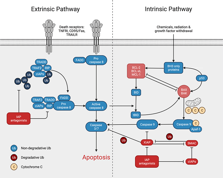
Figure: Extrinsic and intrinsic apoptosis signaling pathway. Proteins and modulators that promote apoptotic cell death are shown in blue, inhibitors are shown in red. (Created with BioRender.com)
New methods for immunogen production, animal immunization, and antibody generation, purification, and validation have matured such that it is now possible to generate antibodies against a wide range of target proteins, regardless of their biochemical characteristics or natural abundance. Each step of the antibody development process is completed within our in-house, controlled environment, delivering unsurpassed repeatability in antibody construction. Each component used in our manufacturing process is developed within our facilities, ensuring that each step of the process can be certified and verified multiple times. Our goal is to provide accountability and repeatable test results with each antibody we develop.
Rockland offers a comprehensive variety of antibodies and antibody-based tools for the numerous methods needed in apoptosis and cell survival measurement. Our products are designed and tested to perform in a variety of assays including flow cytometry, immunohistochemistry, Western blot, and immunoprecipitation and are developed to make a difference in ongoing research. We know through our efforts, that future generations will benefit from the discoveries made using Rockland antibody products.
Apoptosis is a genetically programmed process for the elimination of damaged or redundant cells by activation of caspases (aspartate-specific cysteine proteases). The onset of apoptosis is controlled by numerous interrelating processes. Cell stress stimulates pro-apoptotic signaling pathways that activate caspase proteases and cause mitochondrial dysfunction. The 'extrinsic' pathway involves stimulation of members of the tumor necrosis factor (TNF) receptor, such as TNFRI, CD95/Fas, or TRAILR (death receptors), located at the cell surface, by their specific ligands, such as TNF-alpha, FasL or TRAIL, respectively. The 'intrinsic' pathway is activated mainly by non-receptor stimuli, such as DNA damage, ER stress, metabolic stress, UV radiation, or growth-factor deprivation. The central event in the 'intrinsic' pathway is the mitochondrial outer membrane permeabilization, which leads to the release of cytochrome c. These two pathways converge at the level of effector caspases, such as caspase-3 and caspase-7 that cleave their substrates after aspartic acid residues. It is the balance between the pro-apoptotic and pro-survival signals that eventually determines whether cells will undergo apoptosis, survive, or proliferate. TNF family of ligands activates anti-apoptotic or cell-survival signals as well as apoptotic signals. Anti-apoptotic ligands, like cytokines and growth factors, promote the survival, proliferation, and differentiation of cells through different signaling molecules like AKT and p90RSK as well as anti-apoptotic proteins like BCL-2 and BCL-xL. Withdrawal of these cytokines and growth factors leads to cell death.
Featured Apoptosis Antibodies
| Product | Clonality | Reactivity | Applications |
| APAF1 Antibody | Polyclonal | Human, Mouse, Rat | WB, IHC, IF, ELISA |
| APAF1 Antibody | Polyclonal | Human, Mouse, Rat | WB, IHC, ELISA |
| APAF1 Antibody [2E10] | Monoclonal | Human, Mouse, Rat | WB, IHC, ELISA |
| APAF1 Antibody [5E11] | Monoclonal | Human, Mouse, Rat | IHC, ELISA |
| BAK Antibody | Polyclonal | Human, Mouse | WB, IHC, ELISA |
| BAX Antibody | Monoclonal | Human | IHC |
| BCL2 Antibody | Polyclonal | Human, Mouse | WB, IHC, IF, ELISA |
| BCL2 Antibody | Polyclonal | Human, Mouse, Rat, Bovine | WB, ELISA |
| BCL-xL Antibody | Polyclonal | Human | WB, ELISA |
| BID Antibody | Polyclonal | Human, Mouse | WB, IHC, IF, ELISA |
| BID Antibody | Polyclonal | Human, Mouse | WB, ELISA |
| Caspase-3 Antibody | Polyclonal | Human | IHC, ELISA |
| Caspase-3 Antibody | Monoclonal | Human | WB, IHC, IF, FC |
| Caspase-6 Antibody | Polyclonal | Human | WB, IHC, IF, ELISA |
| Caspase-6 Antibody | Polyclonal | Human | WB, IHC, ELISA |
| Caspase-7 Antibody | Polyclonal | Human, Mouse, Rat | WB, IHC, ELISA |
| Caspase-7 Antibody | Polyclonal | Human, Mouse, Rat | WB, IHC, ELISA |
| Caspase-8 Antibody | Polyclonal | Human, Mouse, Rat | WB, IHC, IF, ELISA |
| Caspase-8 Antibody | Monoclonal | Human | WB, IHC, IF |
| Caspase-9 Antibody | Polyclonal | Human, Mouse, Rat | WB, ELISA |
| Caspase-9 Antibody | Polyclonal | Human | WB, IHC, IF, ELISA, IP |
| Caspase-10 Antibody | Polyclonal | Human | WB, IHC, IF, ELISA |
| CIAP Antibody | Poolyclonal | Human, Mouse | WB, IHC, IF, ELISA |
| DcR1 Antibody | Polyclonal | Human, Mouse, Rat | WB, IHC, IF, ELISA |
| DcR1 Antibody | Polyclonal | Human, Mouse, Rat | WB, IF, ELISA |
| DcR2 Antibody | Polyclonal | Human, Mouse, Rat | WB, IHC, IF, ELISA |
| DR4 Antibody | Polyclonal | Human | WB, IHC, IF, ELISA |
| Death Receptor 4 Antibody | Polyclonal | Human | WB, IHC, IF, ELISA |
| DR5 Antibody | Polyclonal | Human, Mouse, Rat | WB, IHC, IF, ELISA |
| DR5 Antibody | Polyclonal | Human | WB, IHC, IF, FC |
| Mcl-1 Antibody | Polyclonal | Human | WB, IHC, IF, ELISA |
| RIP1 Antibody | Polyclonal | Human | IHC, ELISA |
| Smac Antibody | Polyclonal | Human, Mouse, Rat | WB, IHC, IF, ELISA, IP |
| TNF p55 Receptor Antibody | Polyclonal | Human, Primate | WB, ELISA |
| TNF p55 Receptor Antibody | Polyclonal | Human | WB, ELISA, IP |
| TRAF2 Antibody | Polyclonal | Human | WB, ELISA |
| TRAF2 Antibody | Polyclonal | Human, Mouse, Rat | WB, IHC, ELISA, IP |
Further Reading
- Bertheloot, D., Latz, E., & Franklin, B. S. (2021). Necroptosis, pyroptosis and apoptosis: an intricate game of cell death. Cellular & molecular immunology, 18(5), 1106–1121.
- Carneiro, B. A., & El-Deiry, W. S. (2020). Targeting apoptosis in cancer therapy. Nature reviews. Clinical oncology, 17(7), 395–417.
- Van Opdenbosch, N., & Lamkanfi, M. (2019). Caspases in Cell Death, Inflammation, and Disease. Immunity, 50(6), 1352–1364.