Although it has long been known that copper has a lethal effect on cells at higher doses, only recently has a possible explanation been found. In the March 2022 issue of Science, Tsevtkov et al. reported a copper-induced cell death associated with lipoylated tricarboxylic acid cycle proteins1. By analogy with other types of cell death, this newly discovered form has been termed cuproptosis.
The researchers used various copper ionophores as tools to shuttle copper ions into the cells in specific concentrations and observe the effects they triggered. By using copper-free as well as copper-containing cell culture media and copper chelators as controls, the relationship between cell death and intracellular copper accumulation was proven. But could it be that the elevated copper concentration triggers an already known form of cell death? To answer this question, the researchers led by Peter Tsvetkov investigated the effect of blocking known signaling pathways using knock-outs and inhibitors. It was clearly shown that cell death triggered by copper ionophores differs in its signaling pathway from apoptosis, necroptosis, pyroptosis, and ferroptosis.
The question of identifying the proteins involved in cuproptosis was subsequently investigated using CRISPR-CAS9 screens. Seven genes were identified that were able to escape copper-induced cell death. Surprisingly, some of them were found to be involved in a rare post-translational modification that has so far been documented for only 5 proteins. This modification, named lipoylation, is characterized by the covalent attachment of lipoamide to lysine residues2. One of the candidates found in the initial screens attracted particular interest as deletion of FDX1 resulted in consistent resistance to cuproptosis. Further immunological experiments demonstrated that the knockout of FDX1 also leads to a complete loss of protein lipoylation, identifying FDX1 as a previously unknown regulator of this pathway. The interesting thing here, of course, is how copper-induced cell death and lipoylation are related. The paper strongly suggests that copper is bound to the lipoyl moiety of lipoylated proteins and leads to the aggregation of those proteins and subsequent HSP70 activation.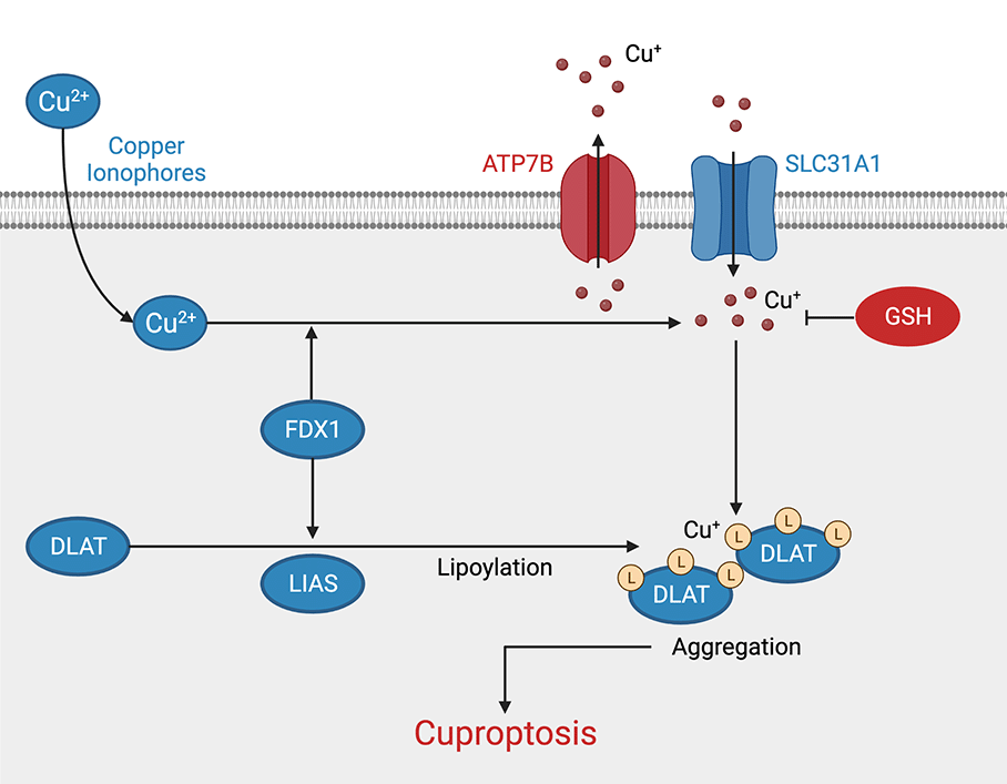
Figure: Cuproptosis signaling pathway. Proteins and modulators that promote copper-induced cell death are shown in blue, inhibitors are shown in red. (Adapted from Tsvetkov et al., 2022, created with BioRender.com)
It remains to be seen whether exploiting these findings can provide new momentum for the research on copper-related disorders, such as Menkes disease, occipital horn syndrome, and Wilson disease, or if it will provide a new approach to treating certain types of tumors.
Cuproptosis Antibodies from antibodies-online
| Product | Clonality | Reactivity | Applications |
| ATP7B Antibody | Monoclonal | Human, Mouse, Rat | WB, IHC, IF, IP |
| DLAT Antibody | Polyclonal | Human, Mouse, Rat, Dog | WB, IHC |
| FDX1 Antibody | Polyclonal | Human | WB, IHC |
| GSH Antibody | Polyclonal | Broad | IHC, ELISA |
| Hsp70 Antibody | Monoclonal | Broad | WB, IHC, FC, ELISA |
| LIAS Antibody | Polyclonal | Human | WB, IHC, ELISA |
References
- Tsvetkov, P., Coy, S., Petrova, B., Dreishpoon, M., Verma, A., Abdusamad, M., Rossen, J., Joesch-Cohen, L., Humeidi, R., Spangler, R. D., Eaton, J. K., Frenkel, E., Kocak, M., Corsello, S. M., Lutsenko, S., Kanarek, N., Santagata, S., & Golub, T. R. (2022). Copper induces cell death by targeting lipoylated TCA cycle proteins. Science (New York, N.Y.), 375(6586), 1254–1261.
- Rowland, E. A., Snowden, C. K., & Cristea, I. M. (2018). Protein lipoylation: an evolutionarily conserved metabolic regulator of health and disease. Current opinion in chemical biology, 42, 76–85.