


Datasheet is currently unavailable. Try again or CONTACT US
ATM phospho S1981 Antibody
Mouse Monoclonal 7C10D8 IgG2a
27 References
200-301-500
100 µg
Liquid (sterile filtered)
WB, ELISA, IHC, IF, IP, Multiplex
Human, Mouse
Mouse
Shipping info:
$65.00 to US & $70.00 to Canada for most products. Final costs are calculated at checkout.
Product Details
Anti-ATM Protein Kinase pS1981 (MOUSE) Monoclonal Antibody - 200-301-500
mouse anti-ATM antibody, mouse anti-ATMpS1981 antibody, mouse anti- ATM pS1981 antibody, DKFZp781A0353 antibody, Human phosphatidylinositol 3 kinase homolog antibody, MGC74674 antibody, Serine protein kinase ATM antibody, T cell prolymphocytic leukemia antibody
Mouse
Monoclonal
IgG2a
Target Details
ATM - View All ATM Products
Human, Mouse
Phosphorylation
Conjugated Peptide
This antibody was produced from a synthetic peptide S-L-A-F-E-E-G-Sp-Q-S-T-T-I-S-S corresponding to aa 1974-1988 of human ATM.
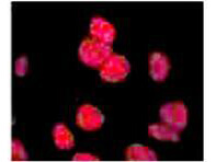
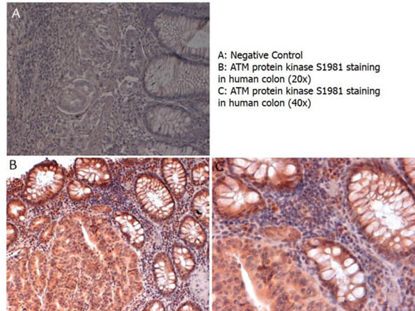
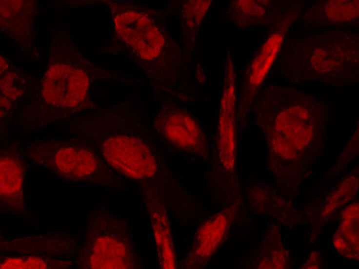
This Protein A Purified Mab antibody is directed against human ATM and is useful in determining its presence by immunohistochemistry. This monoclonal anti-ATM antibody recognizes the phosphorylated form of the protein in native and over-expressed proteins found in various tissues and extracts. Reactivity is observed against human and mouse ATM. Cross reactivity with ATM from other mammalian sources has not been tested.
Application Details
ELISA, IHC
IF, IP, WB, Multiplex
- View References
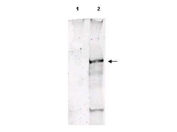
Anti-ATM Protein Kinase pS1981 (MOUSE) Monoclonal Antibody clone has been tested by ELISA and optimized for IHC, but may also be suitable for Western blot, flow cytometry, immunoprecipitation, and immunofluorescence microscopy. Indirect immunoperoxidase staining on formaldehyde-fixed, de-paraffinized tissue sections and/or formalin-fixed cultured cells are recommended when using this antibody as described in Bartkova et al 2005. Product item# 200-301-400 produced from clone 10H11.E12 has been optimized for WB, IP and IF.
Formulation
1.0 mg/mL by UV absorbance at 280 nm
0.02 M Potassium Phosphate, 0.15 M Sodium Chloride, pH 7.2
0.01% (w/v) Sodium Azide
None
Shipping & Handling
Dry Ice
Store vial at -20° C prior to opening. Aliquot contents and freeze at -20° C or below for extended storage. Avoid cycles of freezing and thawing. Centrifuge product if not completely clear after standing at room temperature. This product is stable for several weeks at 4° C as an undiluted liquid. Dilute only prior to immediate use.
Expiration date is one (1) year from date of receipt.
Anti ATM pS1981 Antibody recognizes the product of the ATM gene that is mutated in the hereditary disease ataxia-telangiectasia. ATM codes for a protein kinase that acts as a master regulator of cellular responses to DNA double-strand breaks. ATM is normally inactive and the question of how it is activated in the event of DNA damage (due to ionizing radiation for instance) is central to understanding its function. ATM protein is now shown to be present in undamaged cells as an inactive dimer. Low doses of ionizing radiation, which induce only a few DNA breaks, activate at least half of the total ATM protein present, possibly in response to changes in chromatin structure. The ATM gene encodes a 370-kDa protein that belongs to the phosphoinositide 3-kinase (PI(3)K) superfamily, but which phosphorylates proteins rather than lipids. The 350-amino-acid kinase domain at the carboxy terminus of this large protein is the only segment of ATM with an assigned function. Exposure of cells to IR triggers ATM kinase activity, and this function is required for arrests in G1, S and G2 phases of the cell cycle. Several substrates of the ATM kinase participate in these IR-induced cell-cycle arrests. These include p53, Mdm2 and Chk2 in the G1 checkpoint; Nbs1, Brca1, FancD2 and SMC1 in the transient IR-induced S-phase arrest; and Brca1 and hRad17 in the G2/M checkpoint. See Bakkenist, C. J. & Kastan, M. B. Nature 421, 499-506 (2003) for a complete presentation of this antibody´s specificity and utility.
Altshuller M et al. (2025). ATM promotes reversed fork processing during DNA interstrand cross-link repair. bioRxiv [Preprint].
Applications
WB, IB, PCA
Jiang, Z et al. (2022). Hypophosphorylated pRb knock-in mice exhibit hallmarks of aging and vitamin C-preventable diabetes. The Embo Journal
Applications
IHC, ICC, Histology
Bellusci G et al. (2021). Kinase-independent inhibition of cyclophosphamide-induced pathways protects the ovarian reserve and prolongs fertility. Cell Death Dis.
Applications
IF, Confocal Microscopy
Mattiello L et al. (2021). Asciminib Mitigates DNA Damage Stress Signaling Induced by Cyclophosphamide in the Ovary. Int J Mol Sci.
Applications
IF, Confocal Microscopy
Sakai T et al. (2021). Effects of the Cytoplasm and Mitochondrial Specific Hydroxyl Radical Scavengers TA293 and mitoTA293 in Bleomycin-Induced Pulmonary Fibrosis Model Mice. Antioxidants (Basel).
Applications
WB, IB, PCA
Rosina et al. (2019). Osteogenic differentiation of skeletal muscle progenitor cells is activated by the DNA damage response. Scientific Reports
Applications
WB, IB, PCA
Nagane M et al. (2018). Ataxia-telangiectasia mutated (ATM) kinase regulates eNOS expression and modulates radiosensitivity in endothelial cells exposed to ionizing radiation. Radiat Res.
Applications
IHC, ICC, Histology; WB, IB, PCA
Kim MJ et al. (2018). PAF-Myc-controlled cell stemness is required for intestinal regeneration and tumorigenesis. Dev Cell.
Applications
IHC, ICC, Histology
Emery et al. (2017). Expression and function of ABCG2 and XIAP in glioblastomas. Journal of Neuro-Oncology
Applications
IHC, ICC, Histology
Sinha et al. (2015). A p53/ARF-dependent anticancer barrier activates senescence and blocks tumorigenesis without impacting apoptosis. Molecular Cancer Research
Applications
IF, Confocal Microscopy; IHC, ICC, Histology
Liu et al. (2015). Loss of autophagy causes a synthetic lethal deficiency in DNA repair. Proc. Natl. Acad. Sci. U.S.A
Applications
IF, Confocal Microscopy
Chene G et al. (2015). DNA damage signaling and apoptosis in preinvasive tubal lesions of ovarian carcinoma. Int J Gynecol Cancer.
Applications
IHC, ICC, Histology
Nagane M et al. (2015). Activation of eNOS in endothelial cells exposed to ionizing radiation involves components of the DNA damage response pathway. Biochem Biophys Res Commun.
Applications
WB, IB, PCA
Broering TJ et al. (2014). BRCA1 establishes DNA damage signaling and pericentric heterochromatin of the X chromosome in male meiosis. J Cell Biol
Applications
IF, Confocal Microscopy; IP, Co-IP
Phesse TJ et al. (2014). Endogenous c-Myc is essential for p53-induced apoptosis in response to DNA damage in vivo. Cell Death Differ.
Applications
IHC, ICC, Histology
Shamma A et al. (2013). ATM mediates pRB function to control DNMT1 protein stability and DNA methylation. Mol Cell Biol
Applications
IHC, ICC, Histology; Multiplex Assay
Vidal-Eychenié S, Décaillet C, Basbous J, Constantinou A. (2013). DNA structure-specific priming of ATR activation by DNA-PKcs. J Cell Biol.
Applications
WB, IB, PCA
Schwab KR et al. (2013). Arrested spermatogenesis and evidence for DNA damage in PTIP mutant testes. Dev Biol.
Applications
WB, IB, PCA
Parikh N et al. (2012). Mouse tissues that undergo neoplastic progression after K-Ras activation are distinguished by nuclear translocation of phospho-Erk1/2 and robust tumor suppressor responses. Mol Cancer Res.
Applications
IF, Confocal Microscopy
Raynaud CM et al. (2010). DNA damage repair and telomere length in normal breast, preneoplastic lesions, and invasive cancer. Am J Clin Oncol.
Applications
IF, Confocal Microscopy
Lantuejoul S et al. (2010). Telomere maintenance and DNA damage responses during lung carcinogenesis. Clin Cancer Res.
Applications
IHC, ICC, Histology
Raynaud CM et al. (2009). Telomere length, telomeric proteins and DNA damage repair proteins are differentially expressed between primary lung tumors and their adrenal metastases. Lung Cancer.
Applications
IHC, ICC, Histology
Leemput J et al. (2009). ATM localization and gene expression in the adult mouse eye. Mol Vis.
Applications
IHC, ICC, Histology; WB, IB, PCA
Navaraj A et al. (2009). Reduced cell death, invasive and angiogenic features conferred by BRCA1-deficiency in mammary epithelial cells transformed with H-Ras. Cancer Biol Ther.
Applications
IF, Confocal Microscopy
Raynaud CM et al. (2008). Telomere shortening is correlated with the DNA damage response and telomeric protein down-regulation in colorectal preneoplastic lesions Ann Oncol.
Applications
IHC, ICC, Histology
Bartkova J, Bakkenist CJ, Rajpert-De Meyts E, et al. (2005). ATM activation in normal human tissues and testicular cancer. Cell Cycle
Applications
IP, Co-IP; WB, IB, PCA; IF, Confocal Microscopy
Kitagawa et al. (2004). Phosphorylation of SMC1 is a critical downstream event in the ATM-NBS1-BRCA1 pathway. Genes Dev.
Applications
IF, Confocal Microscopy; WB, IB, PCA; Multiplex Assay
This product is for research use only and is not intended for therapeutic or diagnostic applications. Please contact a technical service representative for more information. All products of animal origin manufactured by Rockland Immunochemicals are derived from starting materials of North American origin. Collection was performed in United States Department of Agriculture (USDA) inspected facilities and all materials have been inspected and certified to be free of disease and suitable for exportation. All properties listed are typical characteristics and are not specifications. All suggestions and data are offered in good faith but without guarantee as conditions and methods of use of our products are beyond our control. All claims must be made within 30 days following the date of delivery. The prospective user must determine the suitability of our materials before adopting them on a commercial scale. Suggested uses of our products are not recommendations to use our products in violation of any patent or as a license under any patent of Rockland Immunochemicals, Inc. If you require a commercial license to use this material and do not have one, then return this material, unopened to: Rockland Inc., P.O. BOX 5199, Limerick, Pennsylvania, USA.