Detection of Akt Migration Using STED Nanoscopy
Written/Edited by David Chimento, PhD
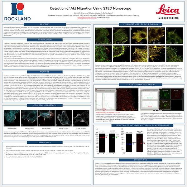
Stimulated Emission Depletion microscopy, or STED microscopy, is a technique that uses the non-linear de-excitation of fluorescent dyes to overcome the resolution limit imposed by diffraction with standard confocal laser scanning microscopes and conventional far-field optical microscopes1. Compared to traditional confocal microscopy, STED offers exceptional improvements in resolution allowing visualization of cellular events at unprecedented levels. Rockland Immunochemicals and Leica Microsystems have jointly analyzed the sub-cellular localization of Akt using both standard confocal microscopy and high-resolution STED nanoscopy. Using resting and EGF-stimulated A431 cells probed with MAb anti-Akt-(p)S473, and PAb anti-Tubulin antibodies, the STED improvement is demonstrated.