

Datasheet is currently unavailable. Try again or CONTACT US
Mouse IgG (H&L) Antibody Pre-adsorbed
Rabbit Polyclonal
10 References
610-4120
2 mg
Liquid (sterile filtered)
ELISA, IF, CUT&RUN, EM
Mouse
Rabbit
Shipping info:
$65.00 to US & $70.00 to Canada for most products. Final costs are calculated at checkout.
Product Details
Rabbit Anti-Mouse IgG (H&L) Antibody (Min X Human Serum Proteins) - 610-4120
rabbit anti-Mouse IgG Antibody, Rabbit-a-Mouse IgG (H&L), Mouse IgG Antibody in rabbit
Rabbit
IgG (H&L)
Polyclonal
IgG
Target Details
Mouse
Native Protein
Anti-Mouse IgG was produced by repeated immunization with mouse IgG whole molecule in rabbit
This product was prepared from monospecific antiserum by immunoaffinity chromatography using Mouse IgG coupled to agarose beads followed by solid phase adsorption(s) to remove any unwanted reactivities. Assay by immunoelectrophoresis (IEP) resulted in a single precipitin arc against anti-Rabbit Serum, Mouse IgG and Mouse Serum. No reaction was observed against Human Serum Proteins.
Application Details
ELISA
EM, IF, CUT&RUN
- View References
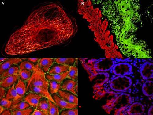
Anti-Mouse IgG (H&L) Antibody has been tested by ELISA and is suitable for immunoblotting (Western or dot blot), ELISA, CUT&RUN, and immunohistochemistry.
Formulation
2.0 mg/mL by UV absorbance at 280 nm
0.02 M Potassium Phosphate, 0.15 M Sodium Chloride, pH 7.2
0.01% (w/v) Sodium Azide
None
Shipping & Handling
Wet Ice
Store vial at 4° C prior to opening. This product is stable for several weeks at 4° C as an undiluted liquid. Dilute only prior to immediate use. For extended storage aliquot contents and freeze at -20° C or below. Avoid cycles of freezing and thawing.
Expiration date is one (1) year from date of receipt.
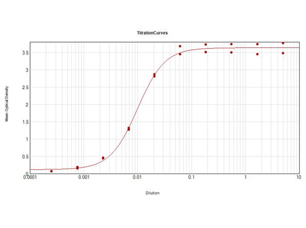
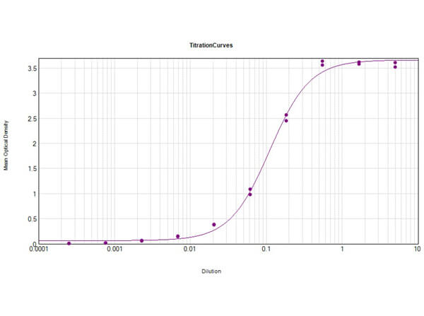
Anti-Mouse IgG antibody generated in rabbit detects specifically mouse IgG (H&L). Secreted as part of the adaptive immune response by plasma B cells, immunoglobulin G constitutes 75% of serum immunoglobulins. Immunoglobulin G binds to viruses, bacteria, as well as fungi and facilitates their destruction or neutralization via agglutination (and thereby immobilizing them), activation of the compliment cascade, and opsonization for phagocytosis. The whole IgG molecule possesses both the F(c) region, recognized by high-affinity Fc receptor proteins, as well as the F(ab) region possessing the epitope-recognition site. Both the Heavy and Light chains of the antibody molecule are present. Secondary Antibodies are available in a variety of formats and conjugate types. When choosing a secondary antibody product, consideration must be given to species and immunoglobulin specificity, conjugate type, fragment and chain specificity, level of cross-reactivity, and host-species source and fragment composition. This anti-Mouse secondary antibody is ideal for investigators who routinely perform titration assays, western-blot, immunoprecipitation and more generally immunoassays.
van der Beek J et al. (2024). Loss of the HOPS complex disrupts early-to-late endosome transition, impairs endosomal recycling and induces accumulation of amphisomes. Mol Biol Cell.
Applications
Immuno-EM
Rawat S et al. (2023). RUFY1 binds Arl8b and mediates endosome-to-TGN CI-M6PR retrieval for cargo sorting to lysosomes. J Cell Biol.
Applications
Immuno-EM
De Mazière A et al. (2022). An optimized protocol for immuno-electron microscopy of endogenous LC3. Autophagy.
Applications
Immuno-gold Electron Microscopy
Burns CH et al. (2021). Pancreatic β-Cell–Specific Deletion of VPS41 Causes Diabetes Due to Defects in Insulin Secretion. Diabetes.
Applications
Immuno-EM
Oorschot V et al. (2021). TEM, SEM, and STEM-based immuno-CLEM workflows offer complementary advantages. Sci Rep.
Applications
TEM
Fujiwara Y et al. (2021). Preparation of optimized concanavalin A-conjugated Dynabeads® magnetic beads for CUT&Tag. PLoS One.
Applications
CUT&RUN
Kooijmans et al. (2018). Recombinant phosphatidylserine-binding nanobodies for targeting of extracellular vesicles to tumor cells: a plug-and-play approach. Nanoscale
Applications
Immuno-EM
Vonk et al. (2018). Mesenchymal Stromal/stem Cell-derived Extracellular Vesicles Promote Human Cartilage Regeneration In Vitro. Theranostics
Applications
Immuno-EM
Kooijmans SAA et al. (2016). PEGylated and targeted extracellular vesicles display enhanced cell specificity and circulation time. J Control Release.
Applications
Immuno-EM
Oorschot VMJ et al. (2014). Immuno correlative light and electron microscopy on Tokuyasu cryosections. Methods Cell Biol.
Applications
IF, Confocal Microscopy; Immuno-EM
This product is for research use only and is not intended for therapeutic or diagnostic applications. Please contact a technical service representative for more information. All products of animal origin manufactured by Rockland Immunochemicals are derived from starting materials of North American origin. Collection was performed in United States Department of Agriculture (USDA) inspected facilities and all materials have been inspected and certified to be free of disease and suitable for exportation. All properties listed are typical characteristics and are not specifications. All suggestions and data are offered in good faith but without guarantee as conditions and methods of use of our products are beyond our control. All claims must be made within 30 days following the date of delivery. The prospective user must determine the suitability of our materials before adopting them on a commercial scale. Suggested uses of our products are not recommendations to use our products in violation of any patent or as a license under any patent of Rockland Immunochemicals, Inc. If you require a commercial license to use this material and do not have one, then return this material, unopened to: Rockland Inc., P.O. BOX 5199, Limerick, Pennsylvania, USA.