IL-1 Beta Antibody
Rabbit Polyclonal
3 References
210-401-319
100 µg
Lyophilized
WB, IHC, IF
Mouse
Rabbit
Shipping info:
$50.00 to US & $70.00 to Canada for most products. Final costs are calculated at checkout.
Product Details
Anti-Mouse IL-1ß (RABBIT) Antibody - 210-401-319
rabbit anti-IL-1 beta antibody, rabbit anti-IL-1b antibody, rabbit anti-Interleukin-1 beta antibody, IL-1ß, catabolin
Rabbit
Polyclonal
IgG
Target Details
Il1b - View All Il1b Products
Mouse
Recombinant Protein
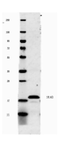
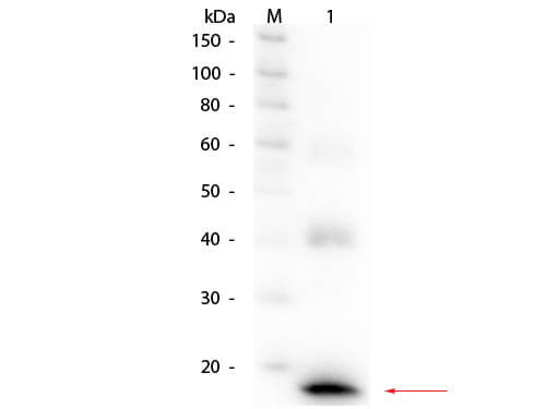
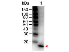
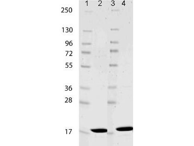
This antibody was prepared by repeated immunizations with recombinant mouse IL-1ß produced in E.coli. The MW of recombinant mouse IL-1ß was 17 kDa.
This is an IgG preparation of whole rabbit serum purified by DEAE fractionation. This antibody is primarily directed against mature, 17,000 MW mouse IL-1ß and is useful in determining its presence in various assays. The antibody does not recognize human IL-1ß or mouse IL-1α based on a neutralization assay. In ELISA formats and other immunoreactive assays, reactivity occurs with rat IL-1ß. This antibody will recognize 10% of the non-denatured (native) precursor 31,000 MW mouse IL-1ß containing samples but will primarily detect all of the 17,000 MW mature molecule. However, in immunoblot analysis, the usual procedure of heating the sample in SDS with or without reducing agents will facilitate denaturing of the 31,000 MW IL- 1ß precursor molecule. Denatured 31,000 precursor IL-1ß will be recognized by this antibody.
Application Details
IF, IHC, WB
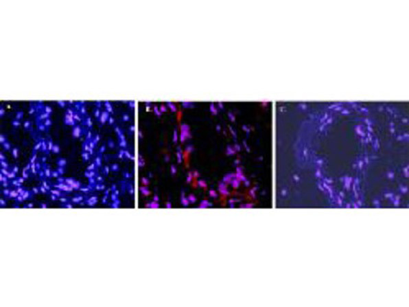
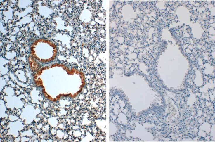
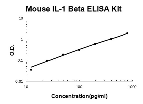
Anti-Mouse IL-1ß has been tested for use in immunohistochemistry, immunoblotting and immunofluorescence. This antibody is useful in ELISA, neutralizations, radioimmunoassays, flow cytometry, and immunoprecipitation. It recognizes the 17,000 MW mature IL-1ß. For immunoblots, typically, IL-1ß is detected from supernatants or lysates of 2 x 10E6 endotoxin-stimulated peripheral blood mononuclear cells (PBMC). PBMC are stimulated for 24 hours with 1% (v/v) serum plus 10 ng/mL E.coli LPS. For immunoprecipitation pre-clearing the preparation with a non-specific Rabbit IgG (p/n 011-001-297) to reduce background is suggested. For immunohistochemistry either paraffin fixation or cryofixation can be used for sample preparation to stain intracellular IL-1ß. For ELISA use HRP Conjugated Anti-Rabbit IgG [H&L] (Goat) (611-1302) for detection. In ELISA formats this antibody is best used as the second antibody in combination with a monoclonal antibody as a capture antibody. This antibody is also useful for neutralization of mouse and rat IL-1ß activity in bioassays. It does not neutralize the biological activity IL-1α. It does not neutralize the biological activity of human or primate IL-1ß. For neutralization, it is recommended to incubate the sample with a dilution of the antibody for at least 4 hours before being tested. A control of similarly diluted normal rabbit IgG is recommended. This antibody can be used for FACS analysis. Caution should be exhibited as the F(c) domain of the rabbit IgG molecule may interact with cells non-specifically.
Formulation
1.0 mg/mL by UV absorbance at 280 nm
0.02 M Potassium Phosphate, 0.15 M Sodium Chloride, pH 7.2
None
None
100 µL
Restore with deionized water (or equivalent)
Shipping & Handling
Ambient
Store Anti-IL-1 beta antibody at 4° C prior to restoration. For extended storage aliquot contents and freeze at -20° C or below. Avoid cycles of freezing and thawing. Centrifuge product if not completely clear after standing at room temperature. This product is stable for several weeks at 4° C as an undiluted liquid. Dilute only prior to immediate use.
Expiration date is six (6) months from date of receipt.
IL-1 beta (also known as Interleukin-1 beta, IL-1ß and catabolin) is produced by activated macrophages. IL-1 stimulates thymocyte proliferation by inducing IL-2 release, B-cell maturation and proliferation, and fibroblast growth factor activity. IL-1 proteins are involved in the inflammatory response, being identified as endogenous pyrogens, and are reported to stimulate the release of prostaglandin and collagenase from synovial cells. IL-1ß is a monomeric secreted protein that may be released by damaged cells or is secreted by a mechanism differing from that used for other secretory proteins. Anti-IL-1 beta antibody is ideal for investigators involved in Cardiovascular and Immunology research.
Rawji KS et al. (2020). Niacin-mediated rejuvenation of macrophage/microglia enhances remyelination of the aging central nervous system. Acta Neuropathol.
Applications
IF, Confocal Microscopy
Rawji KS et al. (2020). Niacin-mediated rejuvenation of macrophage/microglia enhances remyelination of the aging central nervous system. Acta Neuropathol.
Applications
IF, Confocal Microscopy
Bouvier et al. (2016). High Resolution Dissection of Reactive Glial Nets in Alzheimer's Disease. Scientific Reports
Applications
IF, Confocal Microscopy; WB, IB, PCA
This product is for research use only and is not intended for therapeutic or diagnostic applications. Please contact a technical service representative for more information. All products of animal origin manufactured by Rockland Immunochemicals are derived from starting materials of North American origin. Collection was performed in United States Department of Agriculture (USDA) inspected facilities and all materials have been inspected and certified to be free of disease and suitable for exportation. All properties listed are typical characteristics and are not specifications. All suggestions and data are offered in good faith but without guarantee as conditions and methods of use of our products are beyond our control. All claims must be made within 30 days following the date of delivery. The prospective user must determine the suitability of our materials before adopting them on a commercial scale. Suggested uses of our products are not recommendations to use our products in violation of any patent or as a license under any patent of Rockland Immunochemicals, Inc. If you require a commercial license to use this material and do not have one, then return this material, unopened to: Rockland Inc., P.O. BOX 5199, Limerick, Pennsylvania, USA.

